
Breakthrough in Quantum Imaging: First ever low-temperature nanoscale view of exciton spectra
In a new study, researchers have captured the first-ever nanoscale, low-temperature optical images of exciton spectra, opening new doors to the future of quantum material applications.
--
Excitons are tiny, short-lived particles made when light hits a material and briefly knocks an electron loose, leaving behind a positively charged "hole." These particles behave like a pair and play a key role in how materials respond to light. Such excitons dominate the optical response of the ultra-thin, two-dimensional semiconducting materials probed in this work.
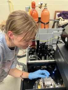
Physics graduate student Anna Roche working on the microscope used in the study.
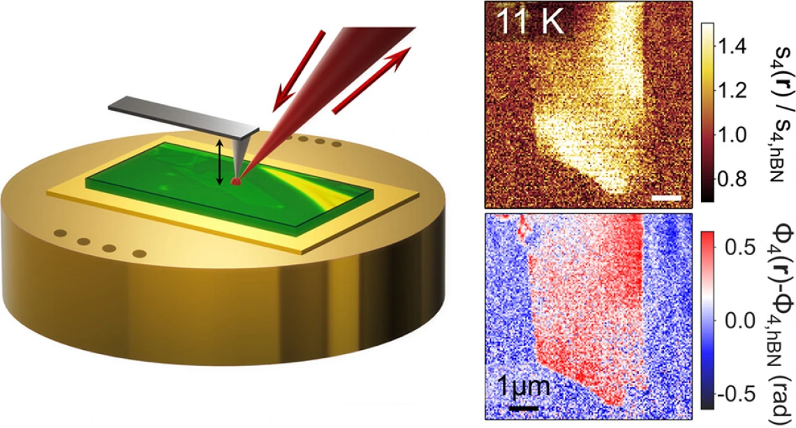
Commonly, scientists study excitonic responses using traditional optical tools that can’t zoom in beyond several hundred nanometers, producing optical images far too blurry to resolve small-scale sample details.
However, in this new study led by University of Arizona Physics Professor Brian LeRoy, Assoc. Professor John Schaibley, and graduate student Anna Roche, researchers used a highly advanced optical microscope that can produce nanoscale optical images and operate at temperatures as low as 10 Kelvin (-441°F). This allowed them to image sample details as small as 50 nanometers — about 2,000 times smaller than the width of a human hair — and detect slight variations in the exciton spectra energy.
The team focused on a material called molybdenum diselenide (MoSe₂) and were able to precisely map how exciton energy varied across its surface. These sharp, detailed images revealed subtle imperfections and variations that were hidden at room temperature, improving scientist’s understanding of sample disorder.
The capabilities demonstrated in this work pave the way for improving the reproducibility and quality of advanced quantum devices.